baby chest x ray pneumonia
Review Of Baby Chest X Ray Pneumonia 2022. Pulmonary consolidation is a non-specific term for pulmonary opacities seen on radiography that are the result from a process that fills the alveolar spaces further discussion here.

Radiographic And Ct Features Of Viral Pneumonia Radiographics
The differential for the radiologic finding of pulmonary consolidation includes blood pulmonary hemorrhage pus infection ie.
. Baby chest x ray pneumonia. Your bronchitis rarely progresses to pneumonia. An x-ray exam will allow your doctor to see your lungs heart and blood vessels to help determine if you have pneumonia.
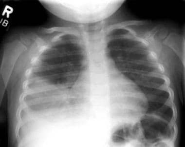
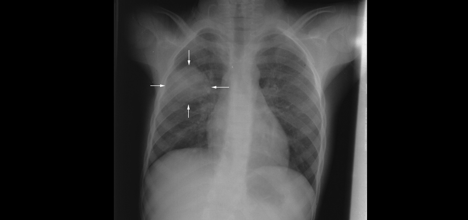
Predictors of the severity of pneumonia have not been thoroughly evaluated among children in developed countries. How to turn on fog lights lincoln mkz. The white spots in the pneumonic X-ray indicated with red arrows called infiltrates distinguish a pneumonic from a healthy condition.
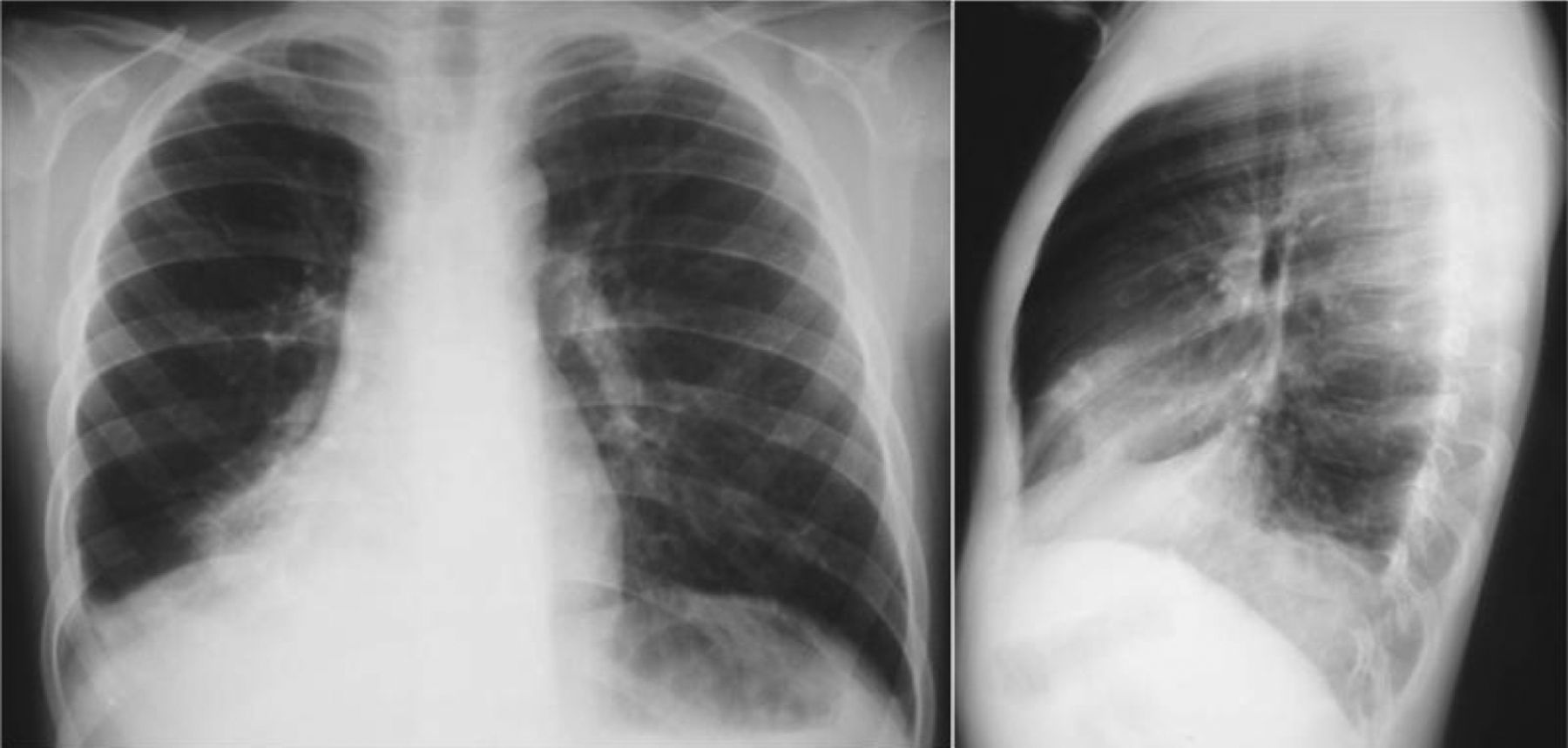
Doctors typically rely on a physical exam and tests. Bacterial pneumonia middle typically exhibits a focal lobar. Megan wright school board.
Largest grain bullet for 500. Pneumatoceles and abscesses are less commonly found but may indicate a Staphylococcus aureus gram-negative or complicated pneumococcal pneumonia. It determines the pathogen that could have caused pneumonia.
Private campgrounds new jersey. A chest X-ray is often used to diagnose pneumonia. Pneumonia remains the most important cause of mortality and morbidity in young children globally 12In addition to the impact of acute disease respiratory infections.
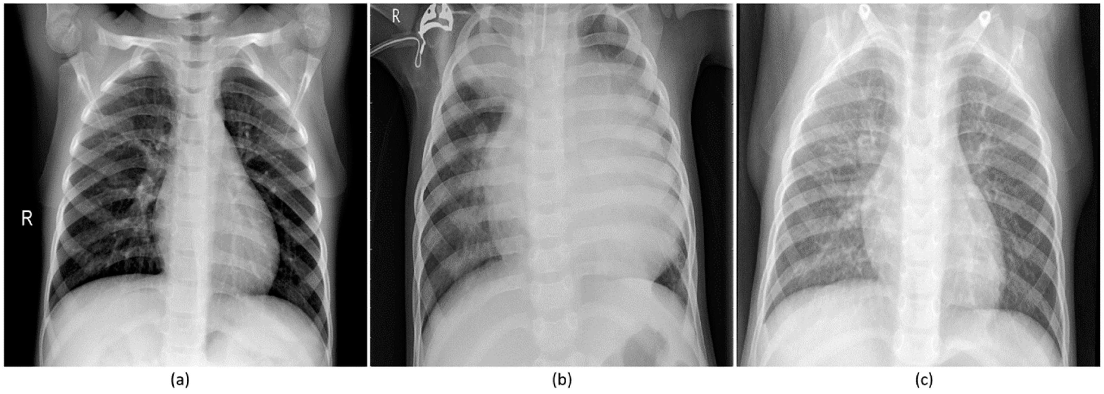
When interpreting the x-ray the radiologist. Jewish baby naming ceremony etiquette. To calculate the incidence and document the clinical features of chest X-ray- CXR- confirmed pneumonia in children aged between 1 month and 5 years living in Greater Suva Fiji.
7 if a child. For this however an x-ray is not helpful as it doesnt provide information for the diagnosis which instead depends on the childs age severity of the symptoms and nature of the infection. When the chest radiograph also includes the abdomen look out for the.
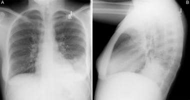
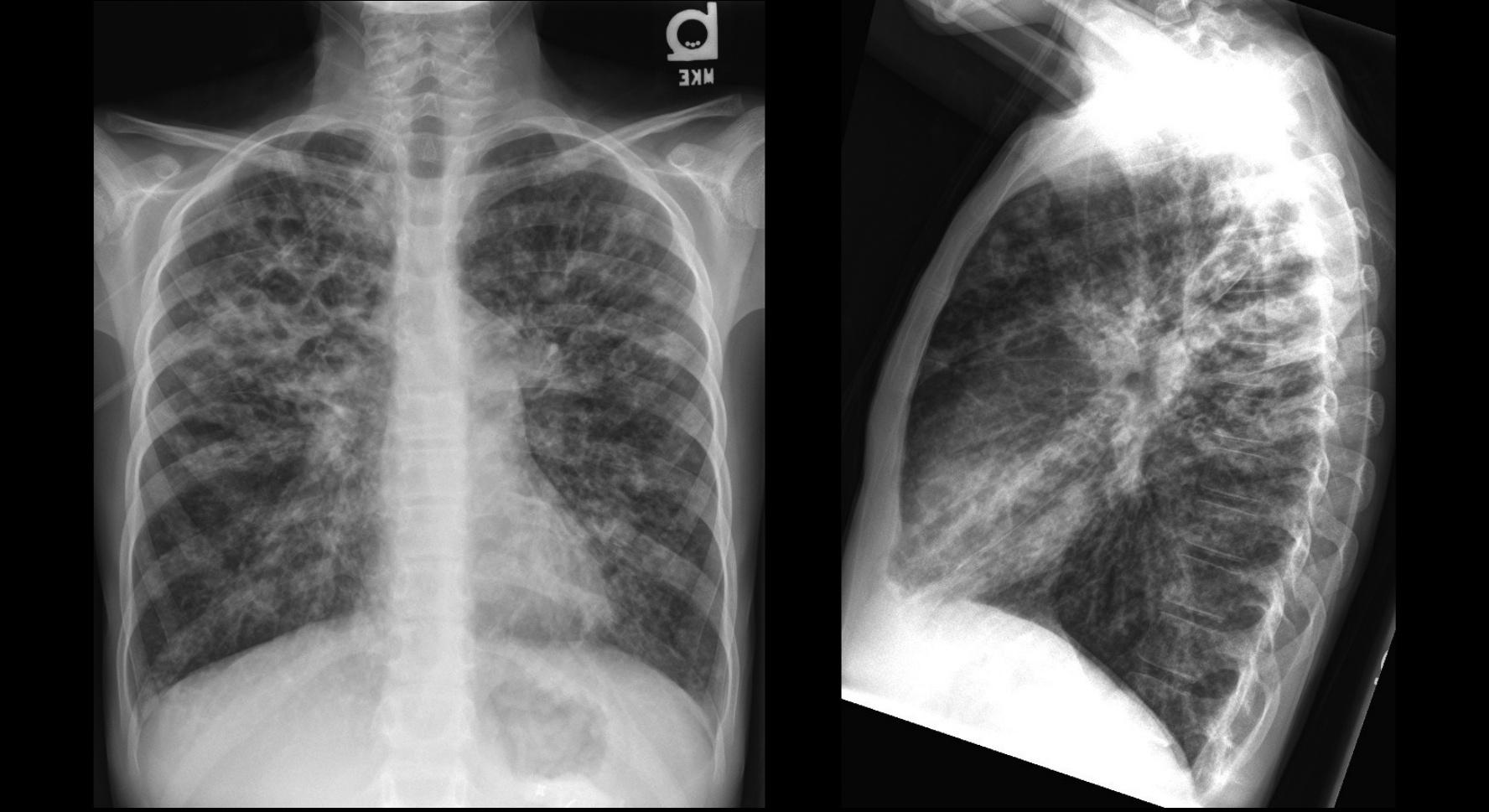
The normal chest X-ray left panel depicts clear lungs without any areas of abnormal opacification in the image. We investigate whether chest radiographic findings could be used as.
Imaging In Pediatric Pneumonia Introduction Chest Radiography Chest Ct Scanning

Complicated Pneumonia In Children The Lancet
A Boy With Recurrent Pneumonia European Respiratory Society
Applied Sciences Free Full Text Visualization And Interpretation Of Convolutional Neural Network Predictions In Detecting Pneumonia In Pediatric Chest Radiographs Html

Pneumonia Imaging Youtube

Chest Imaging Guidance For Pediatric Covid 19 Patients International Consensus
What Can A Chest X Ray Show Cincinnati Children S Blog
What Can A Chest X Ray Show Cincinnati Children S Blog
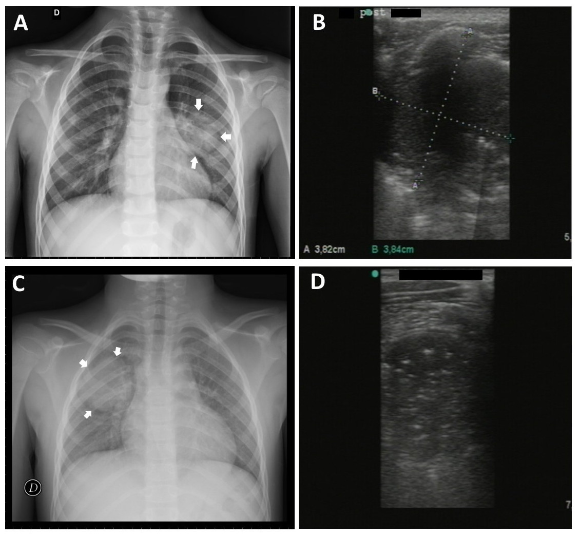
Lung Ultrasound In The Diagnosis Of Pneumonia In Children Proposal For A New Diagnostic Algorithm Peerj
Deep Learning For Classification Of Pediatric Chest Radiographs By Who S Standardized Methodology Plos One
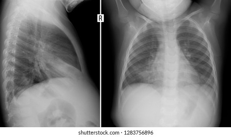
Xray Lungs Child Pneumonia Lung Right Stock Photo 1283756896 Shutterstock
Pneumonia Detection In Chest X Ray Images Using An Ensemble Of Deep Learning Models Plos One

Pediatric Cough Just Another Virus Brown Emergency Medicine

Influenza B Virus Associated Pneumonia In Pediatric Patients Clinical Features Laboratory Data And Chest X Ray Findings Pediatrics Neonatology

Chest Xray Of A Child With A Cough Shows Pneumonia Radiologist Radiology Diagnostic Imaging Medical Imaging X Ray

Round Pneumonia Due To Chlamydia Pneumoniae In A Child Sciencedirect
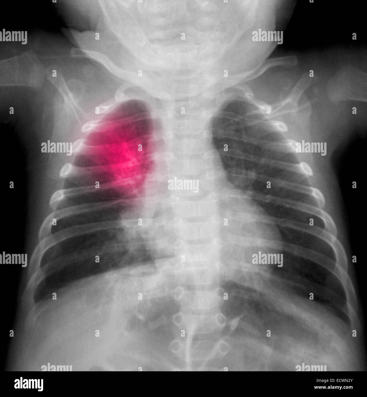
Chest X Ray Of A 3 Month Old Boy Showing Pneumonia Stock Photo Alamy

Diagnosis Of Other Lung Conditions In Premature Babies

Pneumonia X Ray Stock Image C011 9704 Science Photo Library